For information: This matter copy by http://www.geneticacupuncture.com link-
Right and left branches
of large intestine meridian
cross in a very special zone
Right and left branches
of large intestine meridian
cross in a very special zone
| Now we want to
show the reader how other unusual behaviour pattern of an
acupuncture meridian corresponds to an exact anatomic structure.
This is also an exception. In fact, single or multiple crossings
among meridians pertaining to different organs are common (e.g.
the three yin meridians of the lower limb at the point SP-6).
Nevertheless the only meridian that crosses the homologue on the
opposite side of the body is the Large Intestine (LI) meridian.
This crossing takes place at the junction of the upper third and
lower two thirds of the philtrum, or the groove in the upper lip.
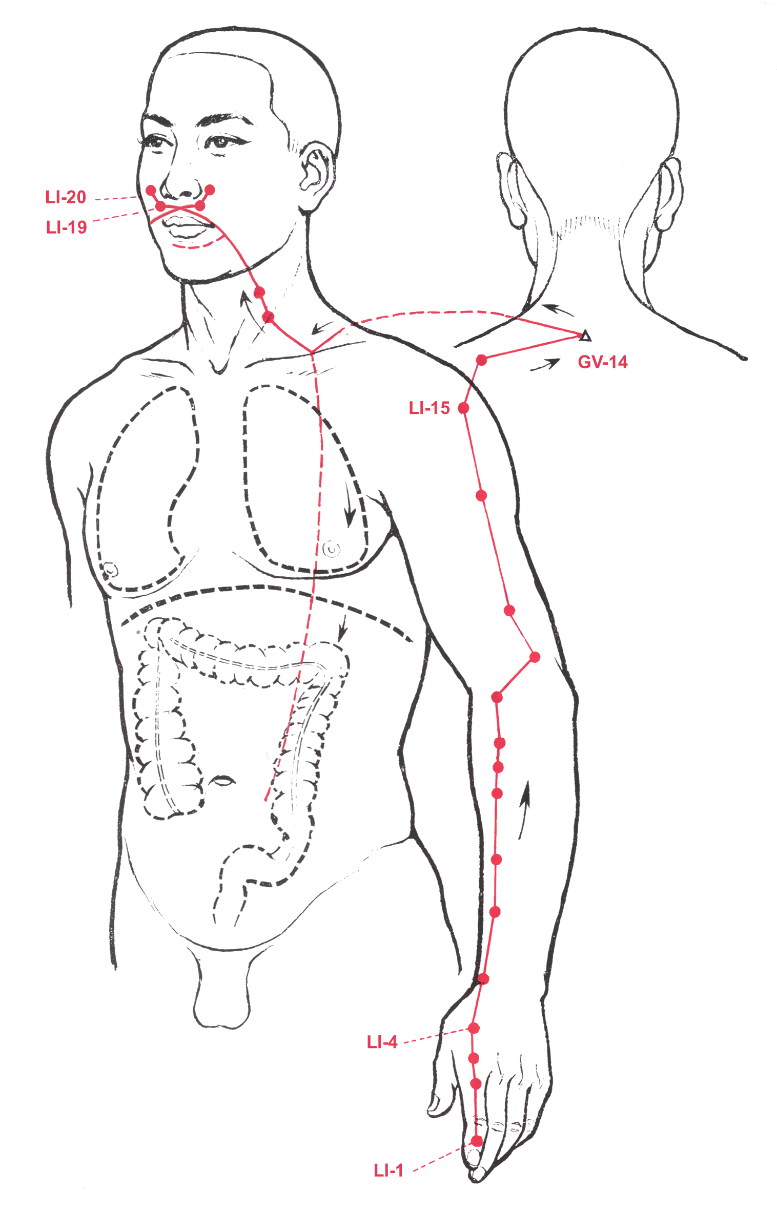
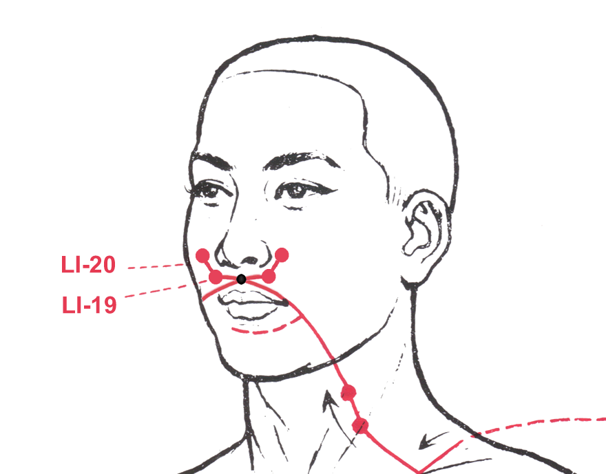
The red line in the picture on the right shows the path and
points of the Large Intestine meridian (the internal path is
dashed). It starts from the tip of the index finger, runs upwards
along the arm and shoulder, goes backwards to the seventh
vertebra, runs upwards and obliquely to the neck and comes
through the jaw. It curves around the upper lip and crosses the
opposite meridian at the philtrum. From here, the left meridian
goes to the right and the right meridian goes to the left, to
either side of the nose, where they end before connecting to the
Stomach (ST) meridian. The point where the two large intestine
meridians cross each other pertains to the Dumai or Governor
Vessel (GV-26), the "extraordinary" meridian (see it here),
that in its superficial path traces out the posterior median
line, from the apex of the coccyx along the spine and skull to the
mucosa above the incisor teeth, where it stops. With the
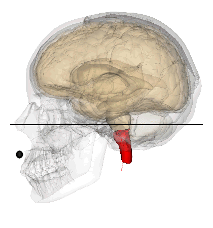
skull in the "Frankfurt plane" position, the philtrum (with the afore-mentioned crossing point) is in correspondence with the decussation of pyramids,
at the level of the second cervical vertebra. Is it just a lucky
combination that the only direct crossing between the right and
left meridian branch of the same organ corresponds to the sole
macroscopically visible crossing of the central nervous system?
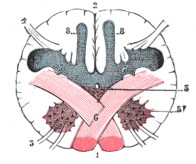
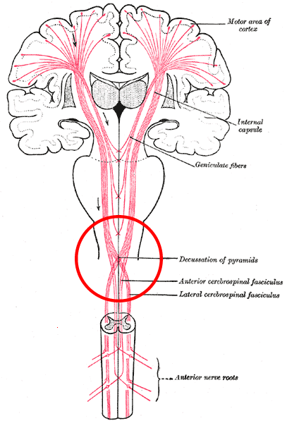
In the picture below you can see the decussation of pyramids on a transverse section.
|  | ||
 | |||
|
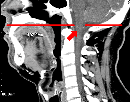
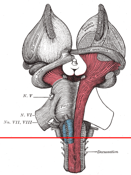
The image on the right is the result of a sagittal CT (X-ray
Computed Tomography), executed on a patient with the skull on the
Frankfurt plane (red line). The arrow indicates where the decussation of pyramids in the medulla oblongata is.
The animated picture below left shows the coincidence on the
sagittal and tranverse planes in the skull between the philtrum (black
circle) and the decussation of pyramids in the brainstem (red).
The black line represents the "Frankfurt plane".
The picture below right shows the details of the crossing
over the philtrum (sutura inter-maxillaris) between the right and
left branches of the large intestine acupuncture meridian.
|  | ||
 |  | ||
| The picture below left simply shows the nerve bundles descending from the cerebral cortex that cross in the decussation of pyramids in the brainstem (red circle). The red horizontal line in the picture on the right underlines the separation between the cranial nerves (non-decussed) and the spinal nerves (partially decussed). As many readers have already learnt elsewhere, the decussation of pyramids is the structure that makes it possible for a unilateral stroke to cause trouble on the same side in the face, and on the opposite side in the limbs. | |||
 |  | ||
|
not to incur
misinterpretation, the decussation of pyramids is visible to naked
eyes, as deducible from the picture above right
| |||
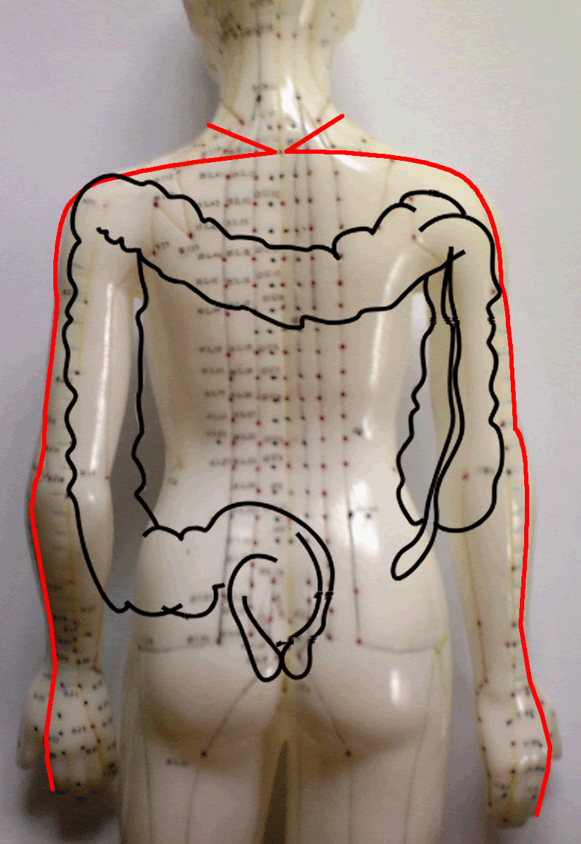
| According to the description given in the Classics (see the first picture above on the right), before passing over the neck and joining the upper lip, where we have seen its right and left branches cross in correspondence with the philtrum, the large intestine meridian begins its pathway from the tip of the index finger, runs over it, over the metacarpal bone, forearm, elbow and arm on the external edge. It surrounds the shoulder joint and from the LI-16 Jugu point, goes backwards to meet the GV-14 Dazhui point of the Governor Vessel (another crossing?), one of the eight Extraordinary meridians, below the 7th cervical spine. Seen from the back, this pathway seems to trace out both the profile of the arms and shoulders and that of the three segments of the large intestine: right, transverse and left colon. Though it is not a double organ like the kidneys, among the abdominal organs the large intestine is the one that has the greatest lateral symmetry and whose two sides are connected (crossed?) via the transverse colon. |
 | ||
|
The point were the two limbs of Large Intestine Channel cross each other (picture below on left) is very special also for embryology, as the philtrum is a privileged site of malformations. By chance, is the cleft palate clinically and genetically associated with large intestine, forefinger, shoulder birth defects? | |||
 |
| ||
|
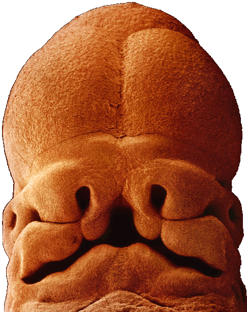
A five
weeks embryo's head. It is nearly 10mm long, and the face is developing,
with mouth, nostrils and eyes. The Large Intestine meridian's right and
left limbs are red, the Governor Vessel black. |
|||
|
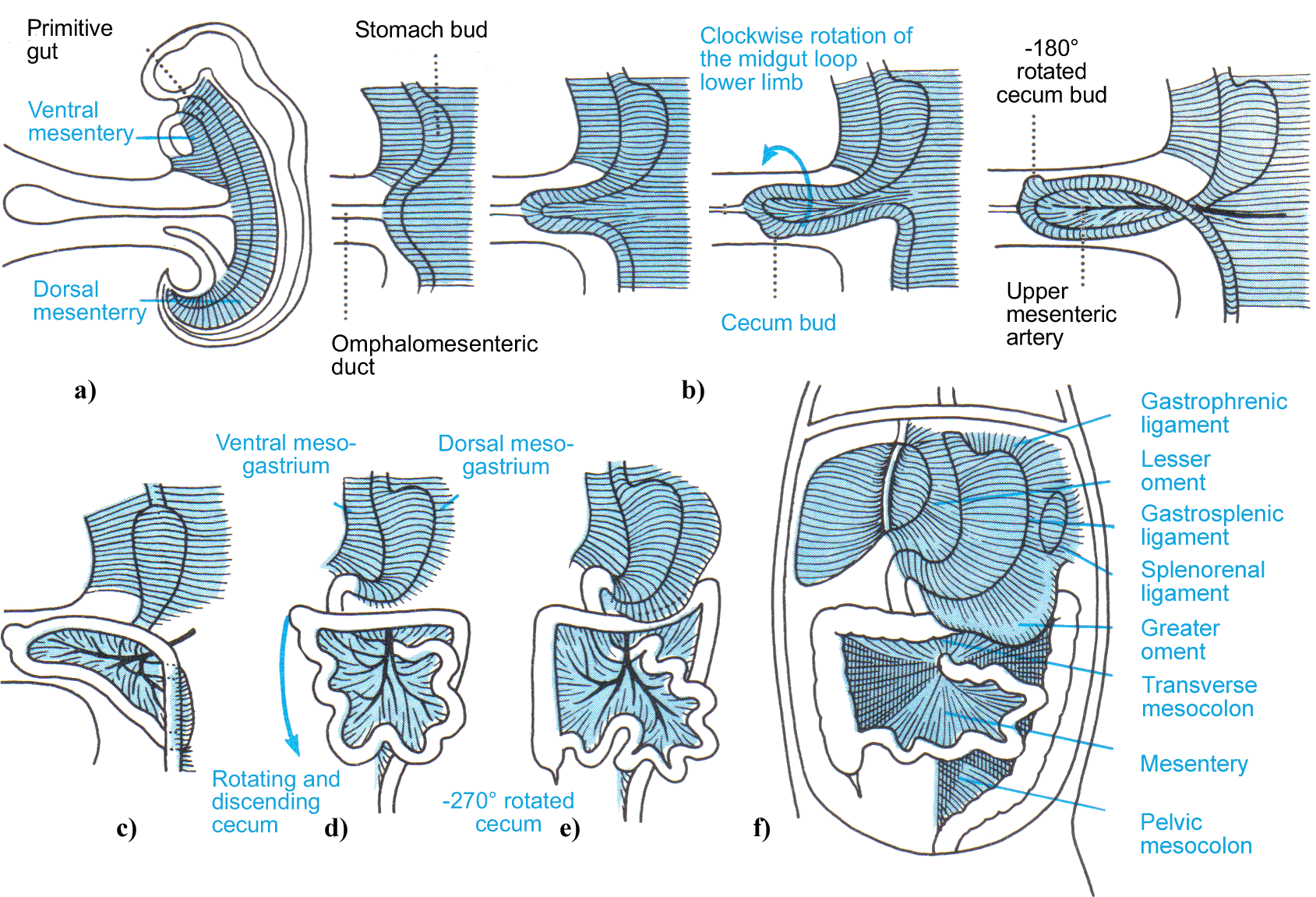
As it is illustrated
in the pictures below, the small and large intestine develop from the
primitive midgut by forming a two-limb loop on the sagittal plane (a), which
detaches itself just below the duodenum that is already taking the shape of
a semicircle opened on the left. The loop herniates physiologically and
temporarily into the umbilical cord (b). Making a 270 degrees global
counterclockwise rotation, the loop first becomes horizontal from vertical
(c), then its lower limb takes progressively the shape of the large
intestine, crosses and overcomes the upper limb (d) that has already begun
folding like an "accordion" to form the small intestine, and rotates itself.
The cecum reaches its definitive position in the right iliac fossa (e), so
that the large intestine circumscribes completely the small intestine (f).
|
|||
 | |||
No comments:
Post a Comment